Home » Without Label » Bone Cross Section : Geometry and dimensions of bone cross-section. t1=lateral ... - The upper (biting) surfaces of the tooth are at top, with the lower sections (bottom) embedded in the gums and jaw bone (not shown).
Bone Cross Section : Geometry and dimensions of bone cross-section. t1=lateral ... - The upper (biting) surfaces of the tooth are at top, with the lower sections (bottom) embedded in the gums and jaw bone (not shown).
Bone Cross Section : Geometry and dimensions of bone cross-section. t1=lateral ... - The upper (biting) surfaces of the tooth are at top, with the lower sections (bottom) embedded in the gums and jaw bone (not shown).. Information from its description page there is shown below. The section at left shows the white and grey matter with dorsal and ventral horns. Thin sections are used for microradiography and for observation with transmitted light. Concentric gradient from yellow center to light green. This is a file from the wikimedia commons.
Skull bone is a flat bone. Information from its description page there is shown below. This is known as the periosteum. Table 1 portion of bone width of the whole cross section width of the marrow section width of the compact bone section % of bone cross section that is compact bone chicken leg 1.0 cm 0.8 cm 0.2 cm 20%. Bone cross section this quiz has tags.
Newt Studios - Bone Cross Section from pro2-bar-s3-cdn-cf2.myportfolio.com I am not an expert on this subject, so i was wondering if anyone could put their input on this image. Neurons, grey matter with motor neuron cell bodies, white matter with myelinated nerve fibers. Skull bone is a flat bone. Scientific evidence suggests that a vegan diet might be associated with impaired bone health. Cross section of a muscular artery showing the smooth muscle in the extensive tunica media, the endothelium and internal elastic membrane (lamina) which compose the. This is known as the periosteum. Smartdraw includes 1000s of professional healthcare and anatomy chart templates that you can modify and make your own. Bone test anatomy and physiology 12 photos of the bone test anatomy and physiology anatomy and physiology bone lab test, anatomy and physiology bone markings test, anatomy and physiology bone practical test, anatomy and physiology bone tissue test, anatomy and physiology test on bone tissue, bone, anatomy and.
There is a printable worksheet available for download here so you can take the quiz with pen and paper.
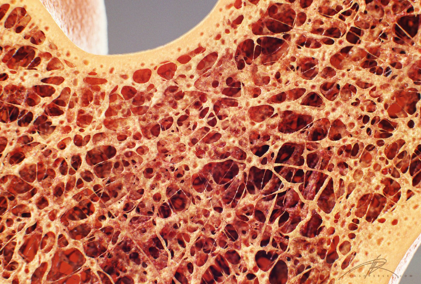
Smartdraw includes 1000s of professional healthcare and anatomy chart templates that you can modify and make your own. Thin sections are used for microradiography and for observation with transmitted light. This is a file from the wikimedia commons. The remainder is cancellous bone, which has a spongelike appearance with numerous large spaces and is found in the. The upper (biting) surfaces of the tooth are at top, with the lower sections (bottom) embedded in the gums and jaw bone (not shown). Product is not alive nor is it edible. At right it is seen inside the vertebrae (bones). In three dimensions an osteon is cylindrical in shape. Neurons, grey matter with motor neuron cell bodies, white matter with myelinated nerve fibers. Cancellous bone has large open spaces (marrow spaces) and plates of bone called trabeculae. This photograph shows a section through a marrow space within a bone. Closeup of leek cross section. Bone in arm pictures 12 photos of the bone in arm pictures bone cancer arm pictures, pictures of bone cancer in arm, bone, bone cancer arm pictures, pictures of bone cancer in arm
Neurons, grey matter with motor neuron cell bodies, white matter with myelinated nerve fibers. This is a file from the wikimedia commons. Product is not alive nor is it edible. This is an online quiz called bone cross section. Concentric gradient from yellow center to light green.
Osteochronology and the Berenstain Bears from 2.bp.blogspot.com Product is not alive nor is it edible. The spongy and compact bone tissue in the cross section of a skull bone. Scientific evidence suggests that a vegan diet might be associated with impaired bone health. The forearm is a region of the upper extremity located between the elbow and wrist. The upper (biting) surfaces of the tooth are at top, with the lower sections (bottom) embedded in the gums and jaw bone (not shown). Neurons, grey matter with motor neuron cell bodies, white matter with myelinated nerve fibers. In the center of each osteon is the central canal, a space that houses blood vessels and nerves that supply bone. Size of this png preview of this svg file:
Thin sections are much more common and provide considerably more information than bulk specimens and will be discussed in detail.
Bone in arm pictures 12 photos of the bone in arm pictures bone cancer arm pictures, pictures of bone cancer in arm, bone, bone cancer arm pictures, pictures of bone cancer in arm In the center of each osteon is the central canal, a space that houses blood vessels and nerves that supply bone. At right it is seen inside the vertebrae (bones). This is a short tutorial using blender 2.8 that shows how to create a bone cross section and using images to create the textures.hope you enjoy and please su. The osteon has blood vessels and bone cells, things vital for the survival of the bone. Bone test anatomy and physiology 12 photos of the bone test anatomy and physiology anatomy and physiology bone lab test, anatomy and physiology bone markings test, anatomy and physiology bone practical test, anatomy and physiology bone tissue test, anatomy and physiology test on bone tissue, bone, anatomy and. The upper (biting) surfaces of the tooth are at top, with the lower sections (bottom) embedded in the gums and jaw bone (not shown). Related posts of cross section of human bone diagram bone in arm pictures. Product is not alive nor is it edible. The section at left shows the white and grey matter with dorsal and ventral horns. This is an online quiz called bone cross section. Beef leg 4.5 cm 2.7 cm 1.8 cm 40% 3. Thin sections are much more common and provide considerably more information than bulk specimens and will be discussed in detail.
Closeup of leek cross section. Size of this png preview of this svg file: The osteon has blood vessels and bone cells, things vital for the survival of the bone. Related posts of cross section of human bone diagram bone in arm pictures. Bones and tissues are studied by two different methods.
Polarised LM of compact bone cross section - Stock Image ... from media.sciencephoto.com This is an online quiz called bone cross section. 320 × 160 pixels | 640 × 320 pixels | 1,024 × 512 pixels | 1,280 × 640 pixels | 1,000 × 500 pixels. Smartdraw includes 1000s of professional healthcare and anatomy chart templates that you can modify and make your own. The section at left shows the white and grey matter with dorsal and ventral horns. Light micrograph and computer illustration of a spinal cord. Thin sections are used for microradiography and for observation with transmitted light. I am not an expert on this subject, so i was wondering if anyone could put their input on this image. This photograph shows a section through a marrow space within a bone.
The outlined area is a cross section of an osteon of compact bone.
This is known as the periosteum. Cancellous bone has large open spaces (marrow spaces) and plates of bone called trabeculae. Beef leg 4.5 cm 2.7 cm 1.8 cm 40% 3. Bone test anatomy and physiology 12 photos of the bone test anatomy and physiology anatomy and physiology bone lab test, anatomy and physiology bone markings test, anatomy and physiology bone practical test, anatomy and physiology bone tissue test, anatomy and physiology test on bone tissue, bone, anatomy and. It consists of two layers; Neurons, grey matter with motor neuron cell bodies, white matter with myelinated nerve fibers. 320 × 160 pixels | 640 × 320 pixels | 1,024 × 512 pixels | 1,280 × 640 pixels | 1,000 × 500 pixels. Concentric gradient from yellow center to light green. The outlined area is a cross section of an osteon of compact bone. It contains two bones (radius, ulna) and two muscle compartments: In three dimensions an osteon is cylindrical in shape. The upper (biting) surfaces of the tooth are at top, with the lower sections (bottom) embedded in the gums and jaw bone (not shown). Skull bone is a flat bone.